

Abstract Motivation The twisted-pair (TP) coil design is a promising strategy for developing novel, flexible, wearable MRI detectors that can provide SNR gains in various clinical applications of high-field MRI. We hypothesize that the TP coil’s receive (Rx) sensitivity can be significantly increased by combining it with two complementary elements, such as dielectric resonators (DRs) and dipole antennas. Methods TP coils were combined with DRs made of high-permittivity material ( ε r = 1070) and transceiver (TxRx) dipole antennas. The Tx and Rx performance of six different types of arrays (TP-only, dipole-only, TP with DRs, dipole with DRs, dipole with TPs, and dipole with TPs and DRs) was investigated through numerical simulations involving a cylindrical phantom suitable for lower extremity applications and two human voxel models. MR phantom experiments were conducted using a 7 Tesla whole-body MRI scanner to validate the Tx and Rx performance of all six array types. Results The array combining all three types of elements (TP coils, DRs, and dipole antennas) provided the highest overall Rx performance; MR phantom experiments showed that integrating DRs with TP coils increased peripheral SNR by 250% and central SNR by 23% (for a total 38% gain in the center when also using dipole antennas in Rx). Human voxel model simulations confirmed that similar SNR gains can be achieved in vivo. Integrating DRs into TP coils also increased central Tx field efficiency by 4.6% and reduced the peak SAR 10g by 25.8% in the human voxel model Hugo. Conclusion DRs and dipole antennas can significantly improve the overall Rx performance of TP coils. This concept can benefit MRI of the human lower extremity at 7 Tesla and encourage exploration of its utility for other clinical applications.
Publication scientifique
Enhancing SNR in MRI at 7T using wearable coils, dielectric resonators, and dipole antennas
Autres publications de la plateforme
Characterising the Diffusion Functional Signature of Negative BOLD With Interleaved TMS...
De Riedmatten, Inès; Spencer, Arthur P. C.; Martuzzi, Roberto; Rochas, Vincent; Pérot, Jean‐Baptiste; Szczepankiewicz, Filip; Jelescu, Ileana O.
Human Brain Mapping
Aberrant cerebrovascular reactivity presents as an early biomarker of psychosis susceptibility...
Delavari, Farnaz; Moia, Stefano; Forrer, Silas; Sandini, Corrado; Kojovic, Nada; Pascucci, Alessandro; Lacour, Clémence; Eliez, Stephan; Van De Ville, Dimitri
Translational Psychiatry
Real-time reinforcement for human-machine interface control
Vassiliadis, Pierre; Pinheiro, Daniel Leal; Fleury, Lisa; Zenon, Alexandre; Esparza-Iaizzo, Martín; Ingster, Abigaïl; Micera, Silvestro; Shokur, Solaiman; Hummel, Friedhelm C.
Neuron
Developmental Alterations in the Diffusion Tensor Imaging Analysis Along the Perivascular...
Pascucci, Alessandro; Forrer, Silas; Sandini, Corrado; Mancini, Valentina; Alemán-Gómez, Yasser; Eliez, Stephan; Delavari, Farnaz
Biological Psychiatry Global Open Science
How Much Does Motion Matter? Evaluating the Motion Robustness of pTx...
Philippe, Jocelyn; Pato Montemayor, Natalia; Romanin, Ludovica; Sleight, Emilie; Klauser, Antoine; Di Noto, Tommaso; Bacha, Lina; Maréchal, Bénédicte; Herrler, Jürgen; Liebig, Patrick A.; Heidemann, Robin M.; Thiran, Jean‐Philippe; Hilbert, Tom; Piredda, Gian Franco; Yu, Thomas
Magnetic Resonance in Medicine
Linguistic pitch is hierarchically encoded in the right ventral stream
Oderbolz, Chantal; Orpella, Joan; Meyer, Martin
Communications Biology
Journal de publication
Magnetic Resonance Materials in Physics, Biology and Medicine
Auteurs:
Wenz, Daniel; Vliem, Jules; Shegurova, Elizaveta; Widmaier, Mark; Xin, Lijing; Karampinos, Dimitrios C.; Zivkovic, Irena
Date de publication:
Plateforme:
Études récentes de la plateforme
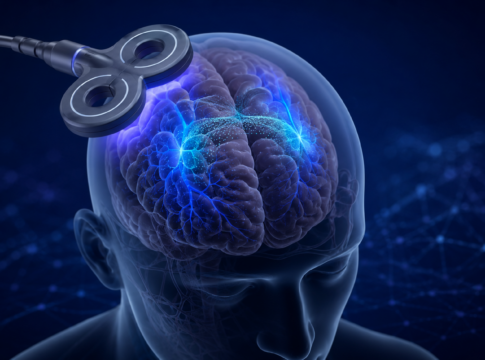
Quand le cerveau s’éteint à l’IRM : un signal plus complexe...
Quand une diminution du signal reste difficile à interpréter
L’imagerie par résonance magnétique fonctionnelle (IRMf) permet de repérer les régions du cerveau qui réagissent...
La connaissance comme récompense
Comparaison de différents types de récompenses
Les participants prennent part à une tâche comprenant trois types de récompenses, de valeur élevée ou faible :
-...
Réseaux cérébraux de la mémoire dans l’épilepsie
Cartographie multimodale des réseaux cérébraux
Combinaison de l'EEG, de l'IRM à champ ultra-élevé et de modèles animaux
Ce projet vise à identifier les marqueurs...