Abstract Stroke patients vary considerably in terms of outcomes: some patients present ‘natural’ recovery proportional to their initial impairment (fitters), while others do not (non-fitters). Thus, a key challenge in stroke rehabilitation is to identify individual recovery potential to make personalized decisions for neuro-rehabilitation, obviating the ‘one-size-fits-all’ approach. This goal requires (i) the prediction of individual courses of recovery in the acute stage; and (ii) an understanding of underlying neuronal network mechanisms. ‘Natural’ recovery is especially variable in severely impaired patients, underscoring the special clinical importance of prediction for this subgroup. Fractional anisotropy connectomes based on individual tractography of 92 patients were analysed 2 weeks after stroke (TA) and their changes to 3 months after stroke (TC − TA). Motor impairment was assessed using the Fugl-Meyer Upper Extremity (FMUE) scale. Support vector machine classifiers were trained to separate patients with natural recovery from patients without natural recovery based on their whole-brain structural connectomes and to define their respective underlying network patterns, focusing on severely impaired patients (FMUE < 20). Prediction accuracies were cross-validated internally, in one independent dataset and generalized in two independent datasets. The initial connectome 2 weeks after stroke was capable of segregating fitters from non-fitters, most importantly among severely impaired patients (TA: accuracy = 0.92, precision = 0.93). Secondary analyses studying recovery-relevant network characteristics based on the selected features revealed (i) relevant differences between networks contributing to recovery at 2 weeks and network changes over time (TC − TA); and (ii) network properties specific to severely impaired patients. Important features included the parietofrontal motor network including the intraparietal sulcus, premotor and primary motor cortices and beyond them also attentional, somatosensory or multimodal areas (e.g. the insula), strongly underscoring the importance of whole-brain connectome analyses for better predicting and understanding recovery from stroke. Computational approaches based on structural connectomes allowed the individual prediction of natural recovery 2 weeks after stroke onset, especially in the difficult to predict group of severely impaired patients, and identified the relevant underlying neuronal networks. This information will permit patients to be stratified into different recovery groups in clinical settings and will pave the way towards personalized precision neurorehabilitative treatment.
Publication scientifique
The structural connectome and motor recovery after stroke: predicting natural recovery
Autres publications de la plateforme
Characterising the Diffusion Functional Signature of Negative BOLD With Interleaved TMS...
De Riedmatten, Inès; Spencer, Arthur P. C.; Martuzzi, Roberto; Rochas, Vincent; Pérot, Jean‐Baptiste; Szczepankiewicz, Filip; Jelescu, Ileana O.
Human Brain Mapping
Aberrant cerebrovascular reactivity presents as an early biomarker of psychosis susceptibility...
Delavari, Farnaz; Moia, Stefano; Forrer, Silas; Sandini, Corrado; Kojovic, Nada; Pascucci, Alessandro; Lacour, Clémence; Eliez, Stephan; Van De Ville, Dimitri
Translational Psychiatry
Real-time reinforcement for human-machine interface control
Vassiliadis, Pierre; Pinheiro, Daniel Leal; Fleury, Lisa; Zenon, Alexandre; Esparza-Iaizzo, Martín; Ingster, Abigaïl; Micera, Silvestro; Shokur, Solaiman; Hummel, Friedhelm C.
Neuron
Developmental Alterations in the Diffusion Tensor Imaging Analysis Along the Perivascular...
Pascucci, Alessandro; Forrer, Silas; Sandini, Corrado; Mancini, Valentina; Alemán-Gómez, Yasser; Eliez, Stephan; Delavari, Farnaz
Biological Psychiatry Global Open Science
How Much Does Motion Matter? Evaluating the Motion Robustness of pTx...
Philippe, Jocelyn; Pato Montemayor, Natalia; Romanin, Ludovica; Sleight, Emilie; Klauser, Antoine; Di Noto, Tommaso; Bacha, Lina; Maréchal, Bénédicte; Herrler, Jürgen; Liebig, Patrick A.; Heidemann, Robin M.; Thiran, Jean‐Philippe; Hilbert, Tom; Piredda, Gian Franco; Yu, Thomas
Magnetic Resonance in Medicine
Linguistic pitch is hierarchically encoded in the right ventral stream
Oderbolz, Chantal; Orpella, Joan; Meyer, Martin
Communications Biology
Journal de publication
Brain
Auteurs:
Koch, Philipp J; Park, Chang-Hyun; Girard, Gabriel; Beanato, Elena; Egger, Philip; Evangelista, Giorgia Giulia; Lee, Jungsoo; Wessel, Maximilian J; Morishita, Takuya; Koch, Giacomo; Thiran, Jean-Philippe; Guggisberg, Adrian G; Rosso, Charlotte; Kim, Yun-Hee; Hummel, Friedhelm C
Date de publication:
Plateforme:
Études récentes de la plateforme
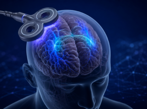
Quand le cerveau s’éteint à l’IRM : un signal plus complexe...
Quand une diminution du signal reste difficile à interpréter
L’imagerie par résonance magnétique fonctionnelle (IRMf) permet de repérer les régions du cerveau qui réagissent...
La connaissance comme récompense
Comparaison de différents types de récompenses
Les participants prennent part à une tâche comprenant trois types de récompenses, de valeur élevée ou faible :
-...
Réseaux cérébraux de la mémoire dans l’épilepsie
Cartographie multimodale des réseaux cérébraux
Combinaison de l'EEG, de l'IRM à champ ultra-élevé et de modèles animaux
Ce projet vise à identifier les marqueurs...