

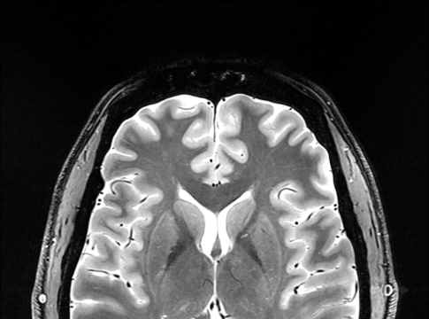
The emergence of 7T clinical MRI technology has sparked our interest in its ability to discern the complex structures of the hand. Our primary objective was to assess the sensory and motor nerve structures of the hand, specifically nerves and Pacinian corpuscles, with the dual purpose of aiding diagnostic endeavors and supporting reconstructive surgical procedures. Ethical approval was obtained to carry out 7T MRI scans on a cohort of volunteers. Four volunteers assumed a prone position, with their hands (N = 8) positioned in a “superman” posture. To immobilize and maintain the hand in a strictly horizontal position, it was affixed to a plastic plate. Passive B0 shimming was implemented. Once high-resolution 3D images had been acquired using a multi-transmit head coil, advanced post-processing techniques were used to meticulously delineate the nerve fiber networks and mechanoreceptors. Across all participants, digital nerves were consistently located on the phalanges area, on average, between 2.5 and 3.5 mm beneath the skin, except within flexion folds where the nerve was approximately 1.8 mm from the surface. On the phalanges area, the mean distance from digital nerves to joints was approximately 1.5 mm. The nerves of the fingers were closer to the bone than to the surface of the skin. Furthermore, Pacinian corpuscles exhibited a notable clustering primarily within the metacarpal zone, situated on the palmar aspect. Our study yielded promising results, successfully reconstructing and meticulously describing the anatomy of nerve fibers spanning from the carpus to the digital nerve division, alongside the identification of Pacinian corpuscles, in four healthy volunteers (eight hands).
Publication scientifique
High-Resolution Magnetic Resonance Neurography at 7T: A Pilot Study of Hand Innervation
Autres publications de la plateforme
Characterising the Diffusion Functional Signature of Negative BOLD With Interleaved TMS...
De Riedmatten, Inès; Spencer, Arthur P. C.; Martuzzi, Roberto; Rochas, Vincent; Pérot, Jean‐Baptiste; Szczepankiewicz, Filip; Jelescu, Ileana O.
Human Brain Mapping
Aberrant cerebrovascular reactivity presents as an early biomarker of psychosis susceptibility...
Delavari, Farnaz; Moia, Stefano; Forrer, Silas; Sandini, Corrado; Kojovic, Nada; Pascucci, Alessandro; Lacour, Clémence; Eliez, Stephan; Van De Ville, Dimitri
Translational Psychiatry
Real-time reinforcement for human-machine interface control
Vassiliadis, Pierre; Pinheiro, Daniel Leal; Fleury, Lisa; Zenon, Alexandre; Esparza-Iaizzo, Martín; Ingster, Abigaïl; Micera, Silvestro; Shokur, Solaiman; Hummel, Friedhelm C.
Neuron
Developmental Alterations in the Diffusion Tensor Imaging Analysis Along the Perivascular...
Pascucci, Alessandro; Forrer, Silas; Sandini, Corrado; Mancini, Valentina; Alemán-Gómez, Yasser; Eliez, Stephan; Delavari, Farnaz
Biological Psychiatry Global Open Science
How Much Does Motion Matter? Evaluating the Motion Robustness of pTx...
Philippe, Jocelyn; Pato Montemayor, Natalia; Romanin, Ludovica; Sleight, Emilie; Klauser, Antoine; Di Noto, Tommaso; Bacha, Lina; Maréchal, Bénédicte; Herrler, Jürgen; Liebig, Patrick A.; Heidemann, Robin M.; Thiran, Jean‐Philippe; Hilbert, Tom; Piredda, Gian Franco; Yu, Thomas
Magnetic Resonance in Medicine
Enhancing SNR in MRI at 7T using wearable coils, dielectric resonators,...
Wenz, Daniel; Vliem, Jules; Shegurova, Elizaveta; Widmaier, Mark; Xin, Lijing; Karampinos, Dimitrios C.; Zivkovic, Irena
Magnetic Resonance Materials in Physics, Biology and Medicine
Journal de publication
Diagnostics
Auteurs:
Guillemin, Pauline C.; Ferreira Branco, David; M’Rad, Yacine; Mattera, Loan; Lorton, Orane; Piredda, Gian Franco; Klauser, Antoine; Martuzzi, Roberto; Poletti, Pierre-Alexandre; Salomir, Rares; Boudabbous, Sana
Date de publication:
Plateforme:
Études récentes de la plateforme
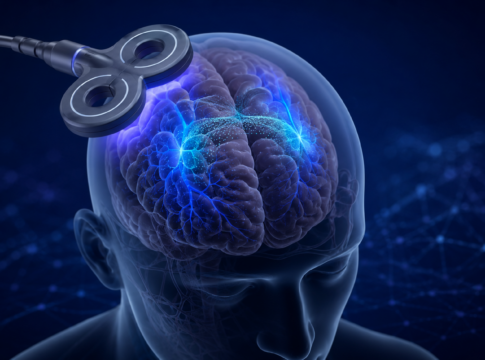
Quand le cerveau s’éteint à l’IRM : un signal plus complexe...
Quand une diminution du signal reste difficile à interpréter
L’imagerie par résonance magnétique fonctionnelle (IRMf) permet de repérer les régions du cerveau qui réagissent...
La connaissance comme récompense
Comparaison de différents types de récompenses
Les participants prennent part à une tâche comprenant trois types de récompenses, de valeur élevée ou faible :
-...
Réseaux cérébraux de la mémoire dans l’épilepsie
Cartographie multimodale des réseaux cérébraux
Combinaison de l'EEG, de l'IRM à champ ultra-élevé et de modèles animaux
Ce projet vise à identifier les marqueurs...