


ABSTRACT The coupling between brain excitatory activity and positive blood oxygen level‐dependent (BOLD) responses is well‐established. Although often associated with inhibition, negative BOLD remains partially understood. Moving away from neurovascular coupling, apparent diffusion coefficient (ADC)‐fMRI provides a more direct measure of excitatory activity, possibly mediated by transient cellular deformations. Diffusion‐weighted fMRI (dfMRI), from which ADC‐fMRI derives, combines vascular and microstructural contributions. While decreases in ADC align with positive BOLD, the possible translation of negative BOLD into positive ADC and the ability of ADC‐fMRI to capture inhibitory activity remain unexplored in humans. In this study, we used transcranial magnetic stimulation (TMS)‐fMRI on the right primary motor cortex (M1) to selectively induce contralateral negative BOLD responses, whilst acquiring interleaved fMRI. TMS was applied at 5 Hz, 90% resting motor threshold, for both BOLD‐fMRI and ADC‐fMRI contrasts, in n = 12 and n = 11 healthy participants, respectively. We replicated previously reported negative BOLD clusters in the contralateral M1 and primary somatosensory cortex (S1). This was accompanied by a negative dfMRI response, but no ADC‐fMRI response, indicating minimal microstructural fluctuations. In ipsilateral M1/S1, no BOLD response was detected while dfMRI revealed a positive cluster, suggesting different sensitivity to the excitatory/inhibitory balance. Overall, combining the findings from BOLD‐fMRI and ADC‐fMRI provides new insights into the vascular and neuronal responses underlying subthreshold TMS and negative BOLD. , Key Points We used interleaved TMS‐fMRI to investigate the diffusion functional signature of negative BOLD. In the region with negative BOLD cluster, a negative diffusion‐weighted fMRI cluster, but no corresponding ADC‐fMRI cluster was detected, suggesting minimal microstructural fluctuations. The absence of a BOLD cluster at the stimulation site, alongside a strong diffusion‐weighted fMRI cluster ( b = 200 s mm −2 ) suggests a different sensitivity to the excitatory/inhibitory balance.
Publication scientifique
Characterising the Diffusion Functional Signature of Negative BOLD With Interleaved TMS ‐ fMRI in the Human Brain
Autres publications de la plateforme
Aberrant cerebrovascular reactivity presents as an early biomarker of psychosis susceptibility...
Delavari, Farnaz; Moia, Stefano; Forrer, Silas; Sandini, Corrado; Kojovic, Nada; Pascucci, Alessandro; Lacour, Clémence; Eliez, Stephan; Van De Ville, Dimitri
Translational Psychiatry
Real-time reinforcement for human-machine interface control
Vassiliadis, Pierre; Pinheiro, Daniel Leal; Fleury, Lisa; Zenon, Alexandre; Esparza-Iaizzo, Martín; Ingster, Abigaïl; Micera, Silvestro; Shokur, Solaiman; Hummel, Friedhelm C.
Neuron
Developmental Alterations in the Diffusion Tensor Imaging Analysis Along the Perivascular...
Pascucci, Alessandro; Forrer, Silas; Sandini, Corrado; Mancini, Valentina; Alemán-Gómez, Yasser; Eliez, Stephan; Delavari, Farnaz
Biological Psychiatry Global Open Science
How Much Does Motion Matter? Evaluating the Motion Robustness of pTx...
Philippe, Jocelyn; Pato Montemayor, Natalia; Romanin, Ludovica; Sleight, Emilie; Klauser, Antoine; Di Noto, Tommaso; Bacha, Lina; Maréchal, Bénédicte; Herrler, Jürgen; Liebig, Patrick A.; Heidemann, Robin M.; Thiran, Jean‐Philippe; Hilbert, Tom; Piredda, Gian Franco; Yu, Thomas
Magnetic Resonance in Medicine
Enhancing SNR in MRI at 7T using wearable coils, dielectric resonators,...
Wenz, Daniel; Vliem, Jules; Shegurova, Elizaveta; Widmaier, Mark; Xin, Lijing; Karampinos, Dimitrios C.; Zivkovic, Irena
Magnetic Resonance Materials in Physics, Biology and Medicine
Linguistic pitch is hierarchically encoded in the right ventral stream
Oderbolz, Chantal; Orpella, Joan; Meyer, Martin
Communications Biology
Journal de publication
Human Brain Mapping
Auteurs:
De Riedmatten, Inès; Spencer, Arthur P. C.; Martuzzi, Roberto; Rochas, Vincent; Pérot, Jean‐Baptiste; Szczepankiewicz, Filip; Jelescu, Ileana O.
Date de publication:
Plateforme:
Études récentes de la plateforme
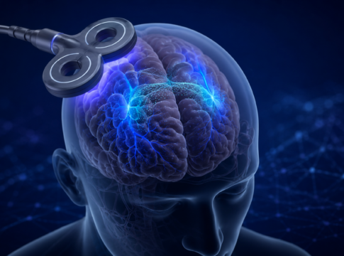
Quand le cerveau s’éteint à l’IRM : un signal plus complexe...
Quand une diminution du signal reste difficile à interpréter
L’imagerie par résonance magnétique fonctionnelle (IRMf) permet de repérer les régions du cerveau qui réagissent...
La connaissance comme récompense
Comparaison de différents types de récompenses
Les participants prennent part à une tâche comprenant trois types de récompenses, de valeur élevée ou faible :
-...
Réseaux cérébraux de la mémoire dans l’épilepsie
Cartographie multimodale des réseaux cérébraux
Combinaison de l'EEG, de l'IRM à champ ultra-élevé et de modèles animaux
Ce projet vise à identifier les marqueurs...